Gallery

Drawn by Fellows.

Work made during the Fellowship — most of it by students who arrived convinced they couldn't draw.

Watch a drawing come together

Time lapses from Fellows' work — press play on any clip.
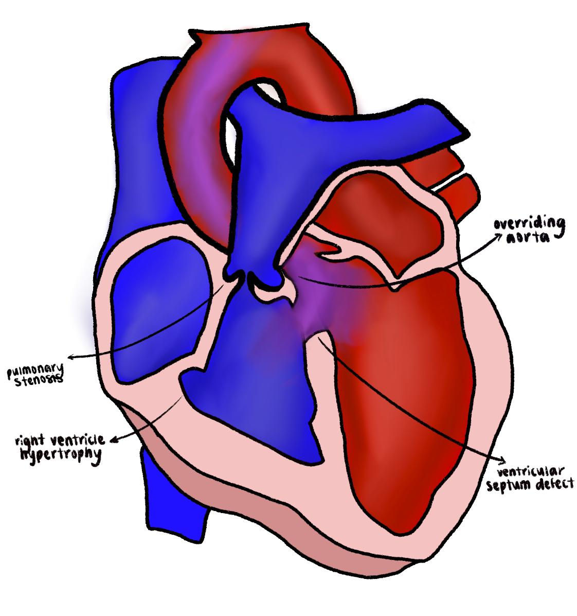
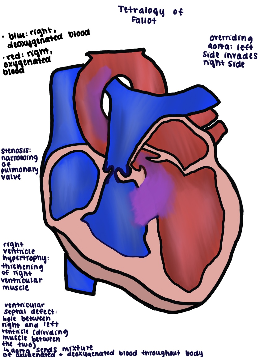
Fellow's drawing of tetralogy of Fallot with labelled overriding aorta, pulmonary stenosis, ventricular septal defect and right ventricular hypertrophy
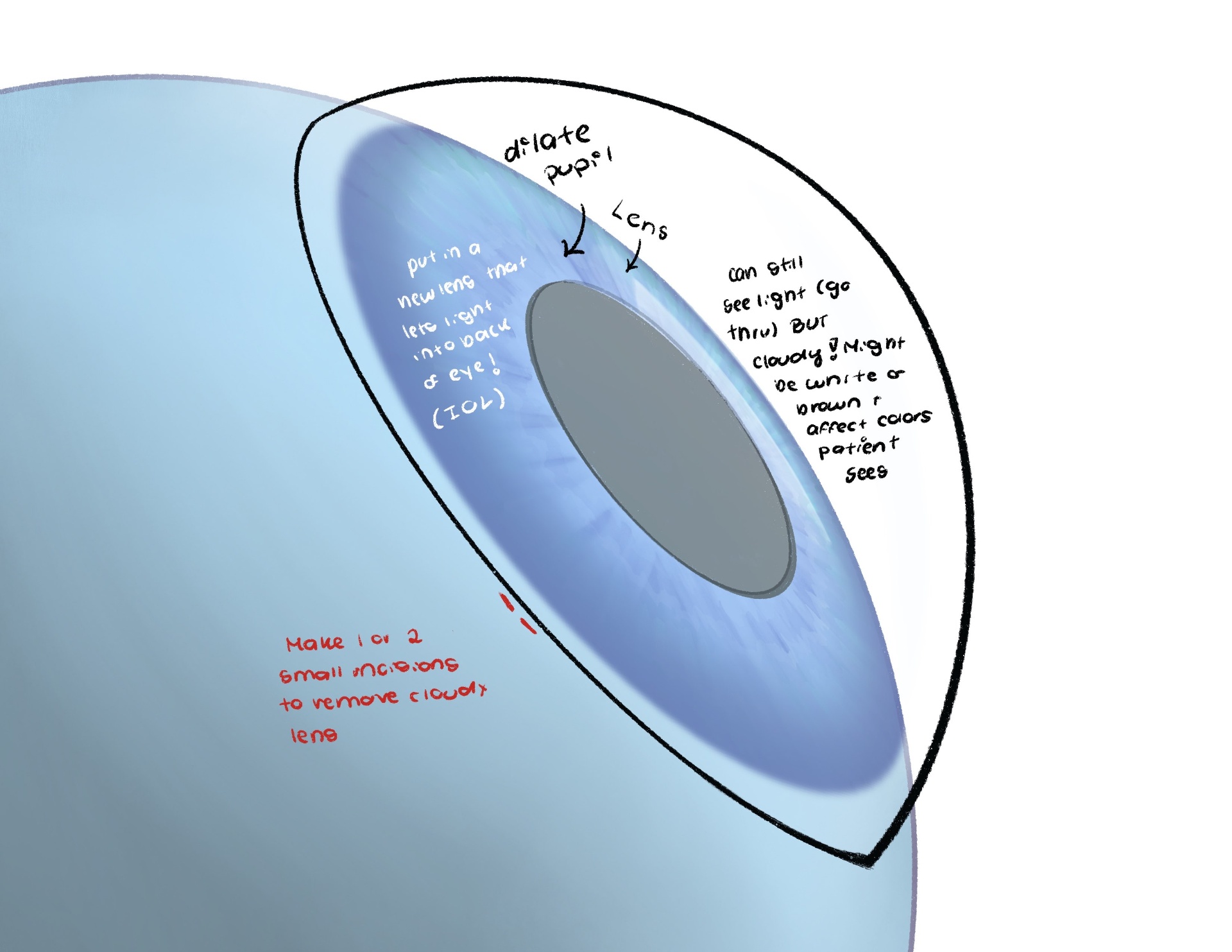
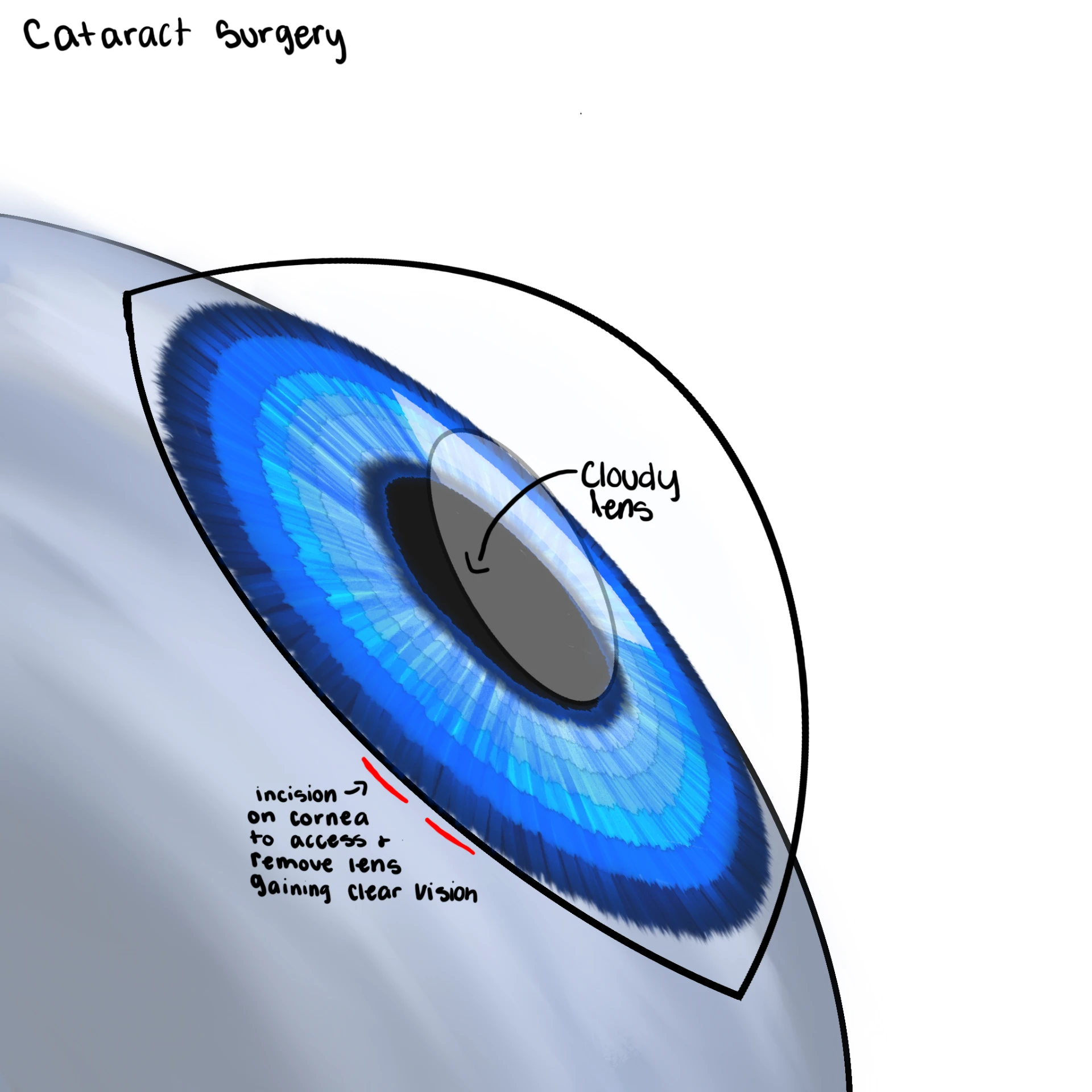
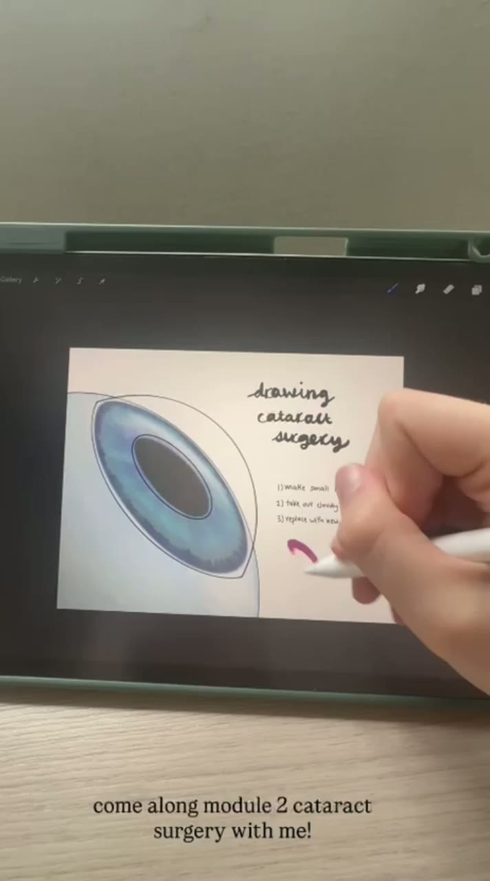
Fellow's annotated drawing of cataract surgery showing the cloudy lens, dilated pupil and lens replacement
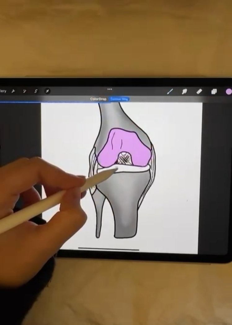
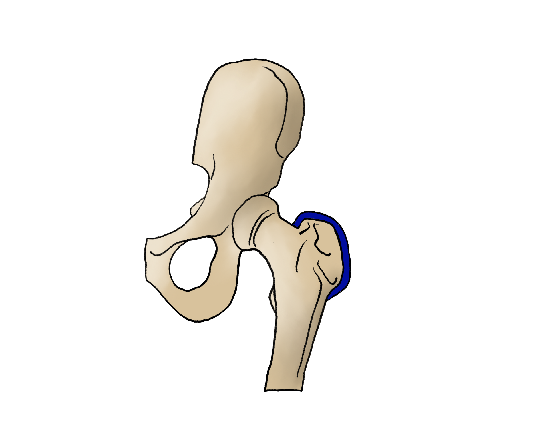
Fellow's drawing of the hip joint with the femoral head cartilage highlighted
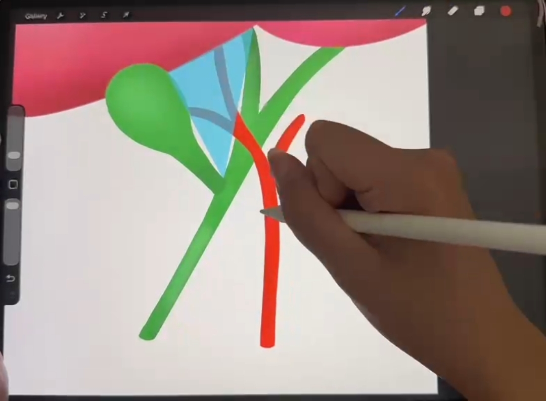
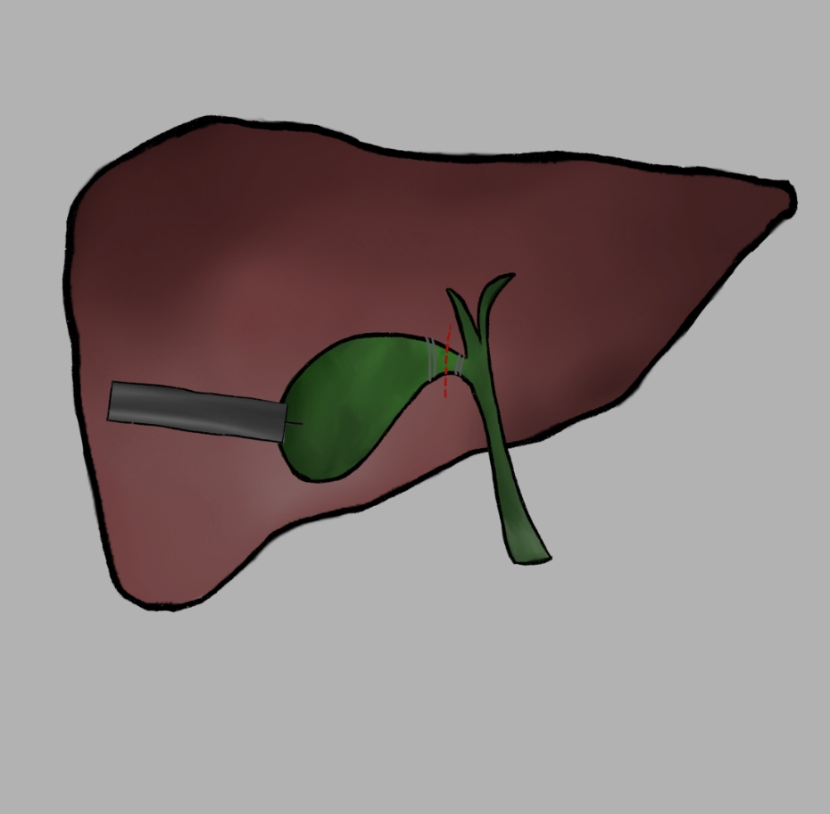
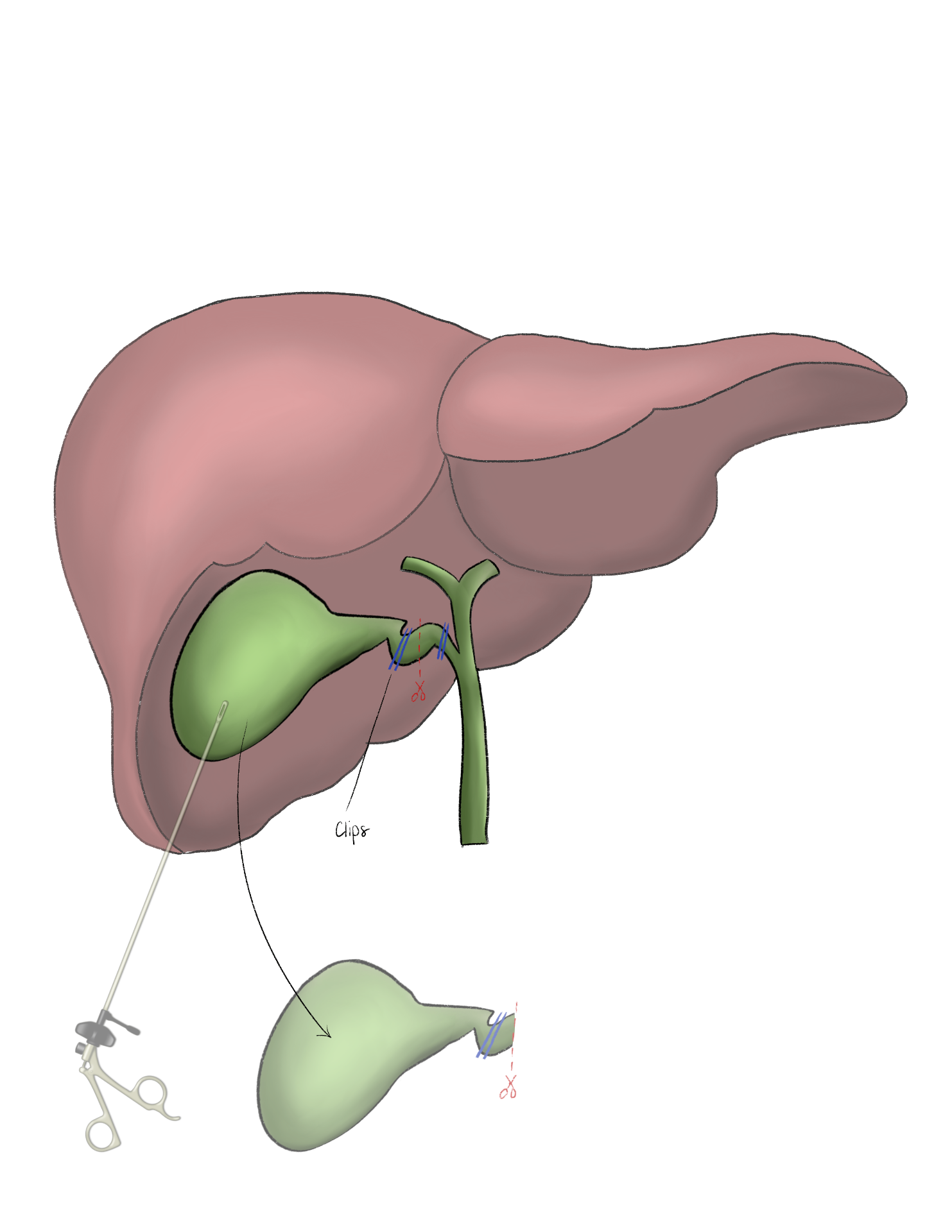
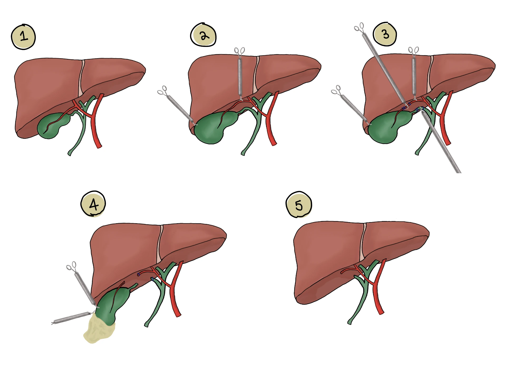
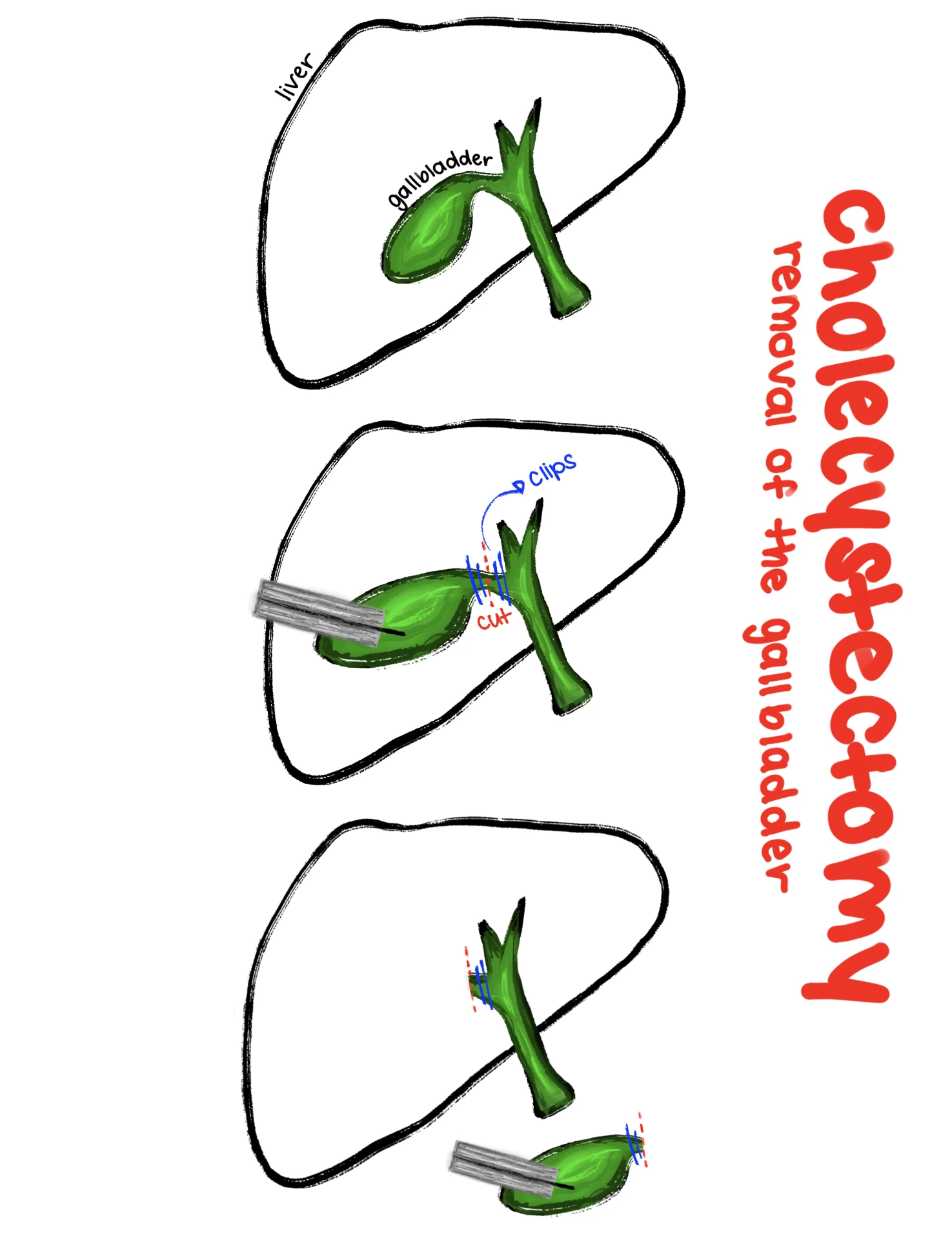
Fellow's drawing of the liver and gallbladder during a laparoscopic cholecystectomy
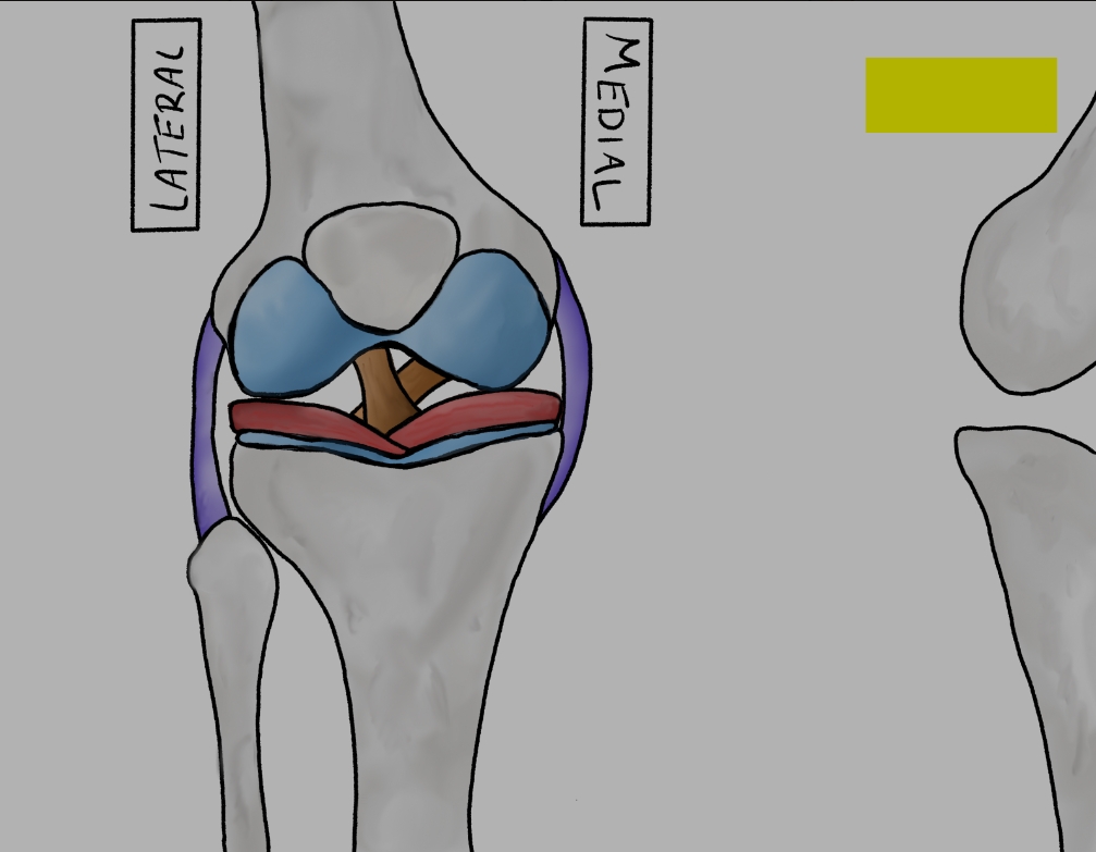
Fellow's drawing of knee joint anatomy with medial and lateral labels, menisci and ligaments
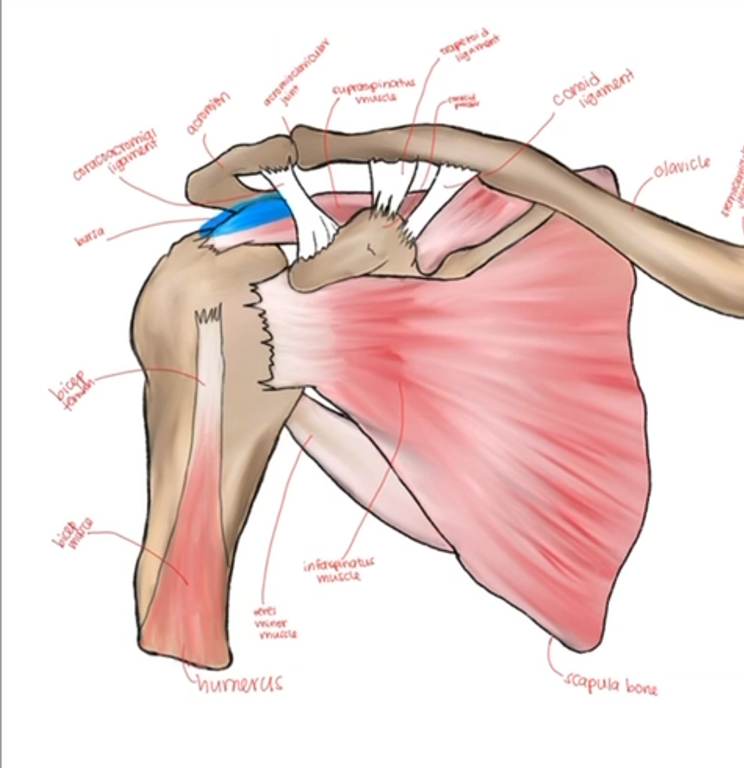
Fellow's fully labelled drawing of shoulder anatomy including rotator cuff muscles, ligaments and bursa
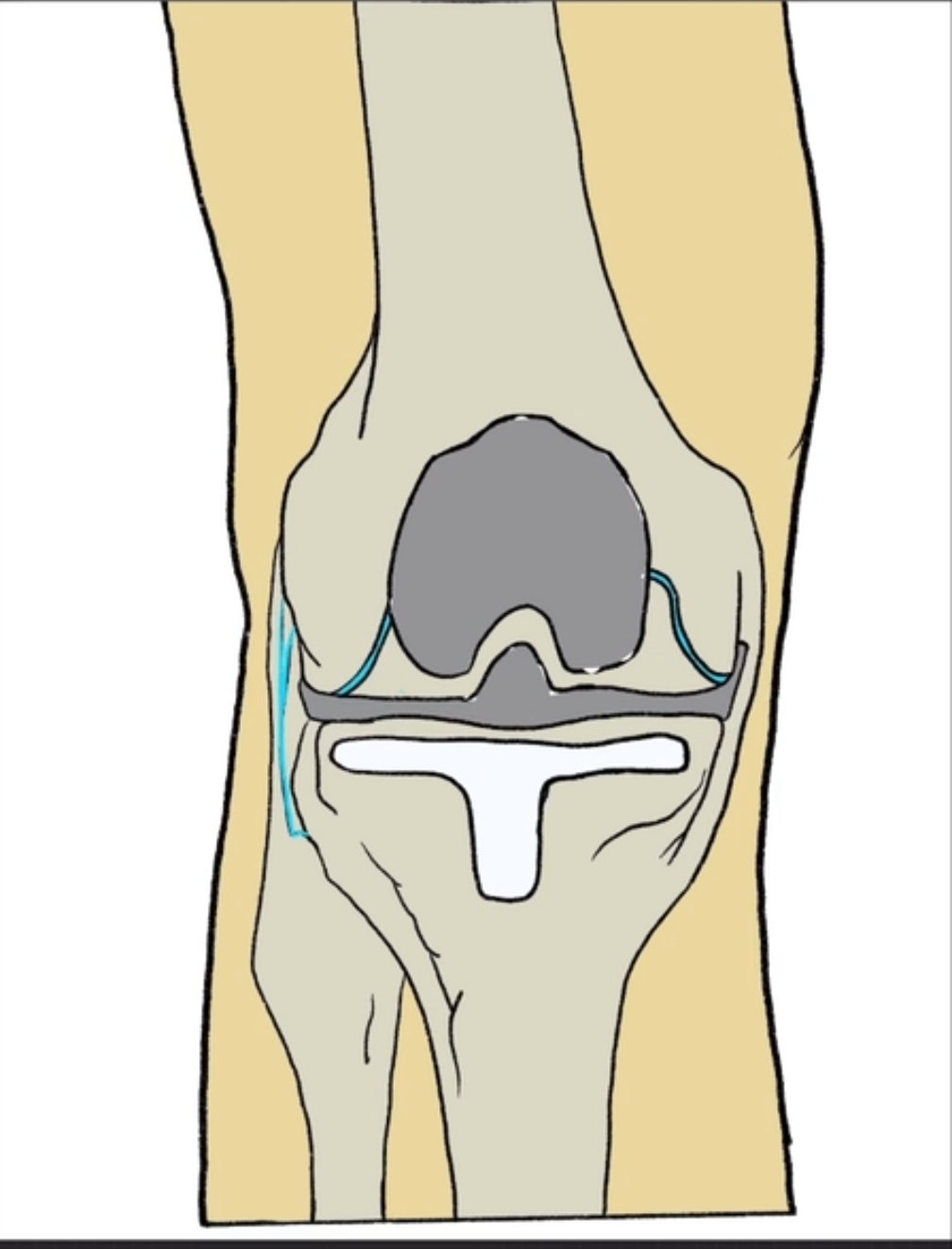
Fellow's drawing of a total knee replacement with implanted femoral and tibial components
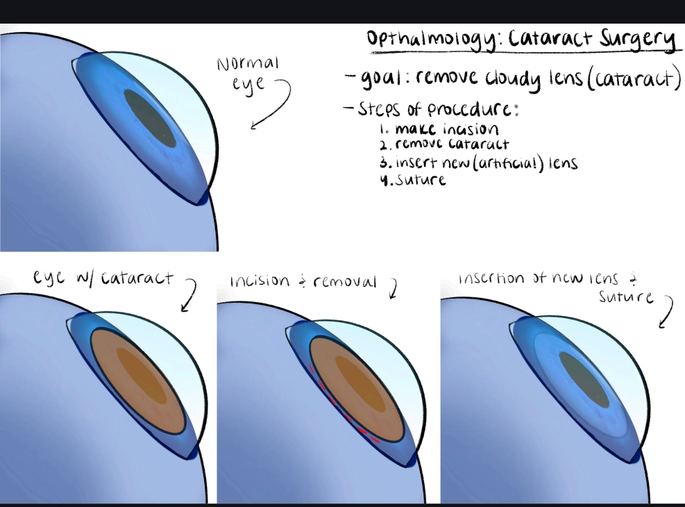
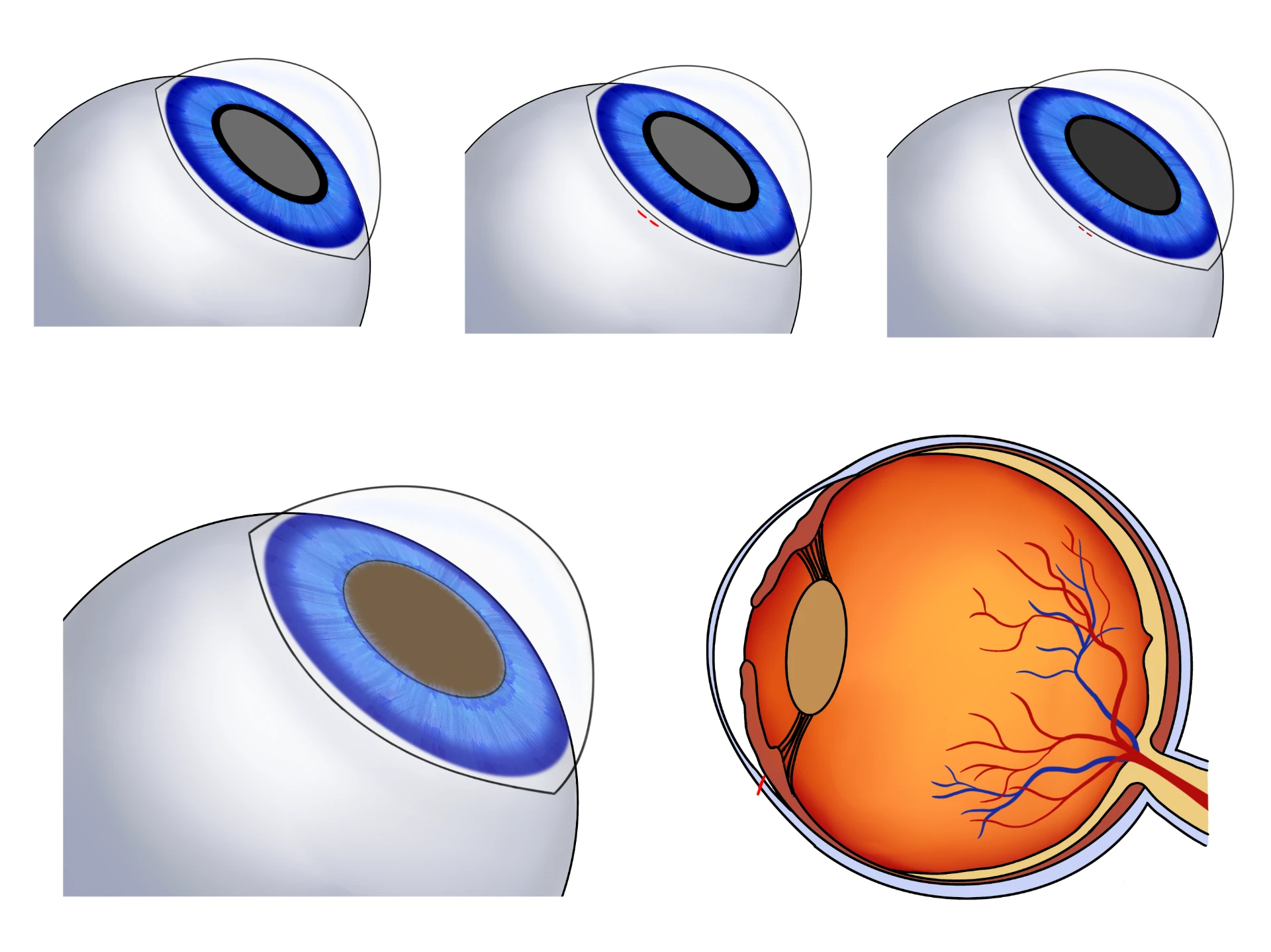
Fellow's four-panel drawing of the steps of cataract surgery from normal eye to new lens insertion
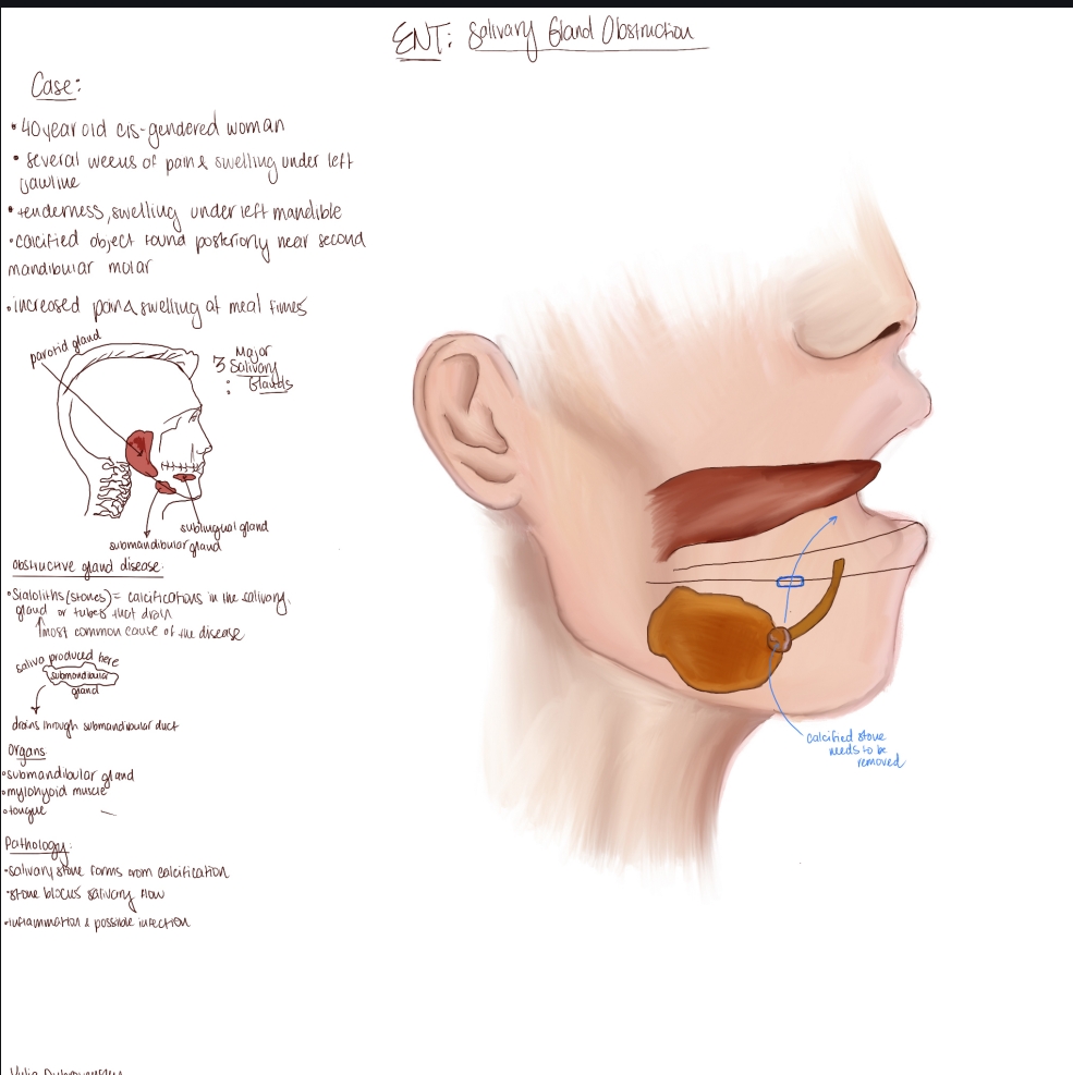
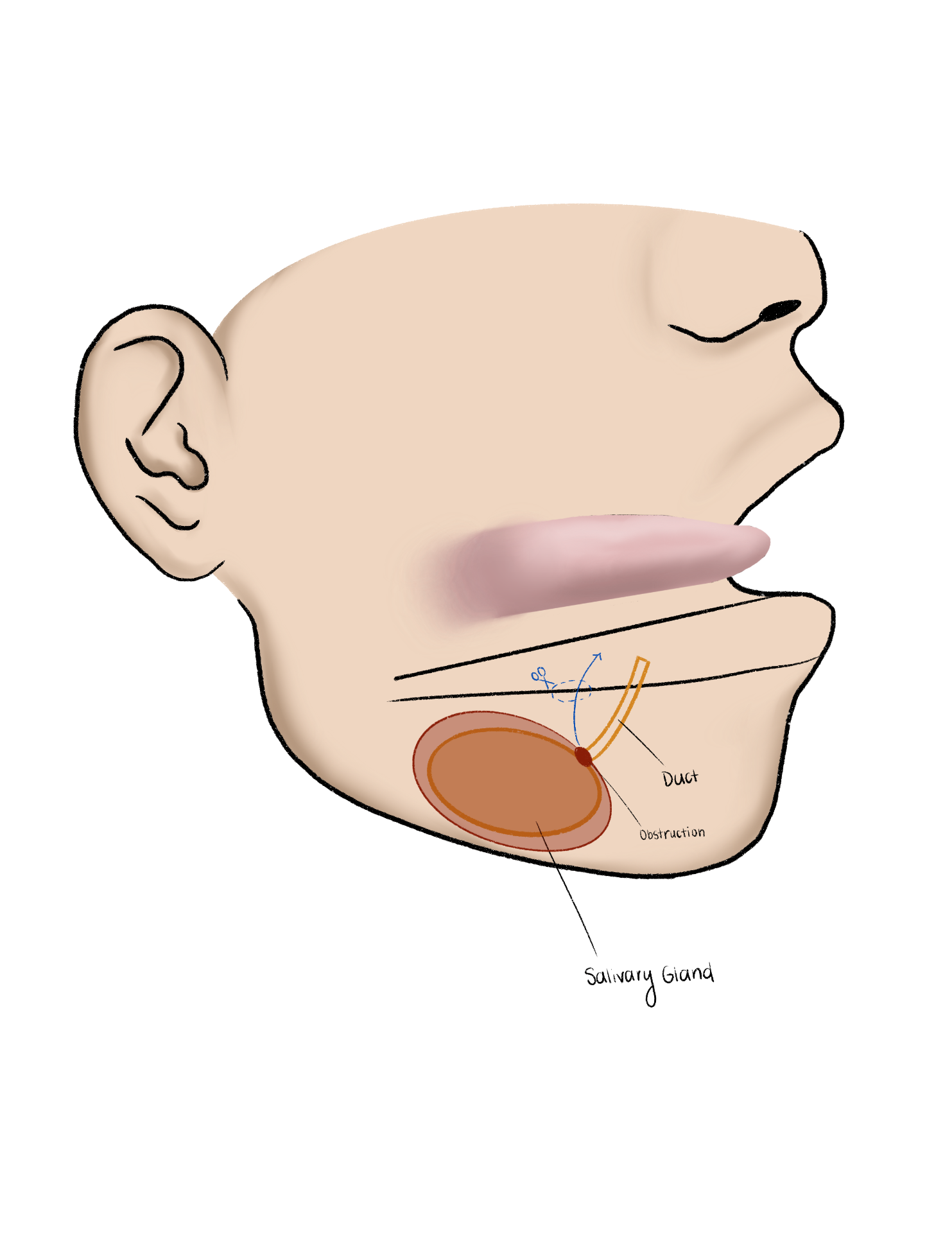
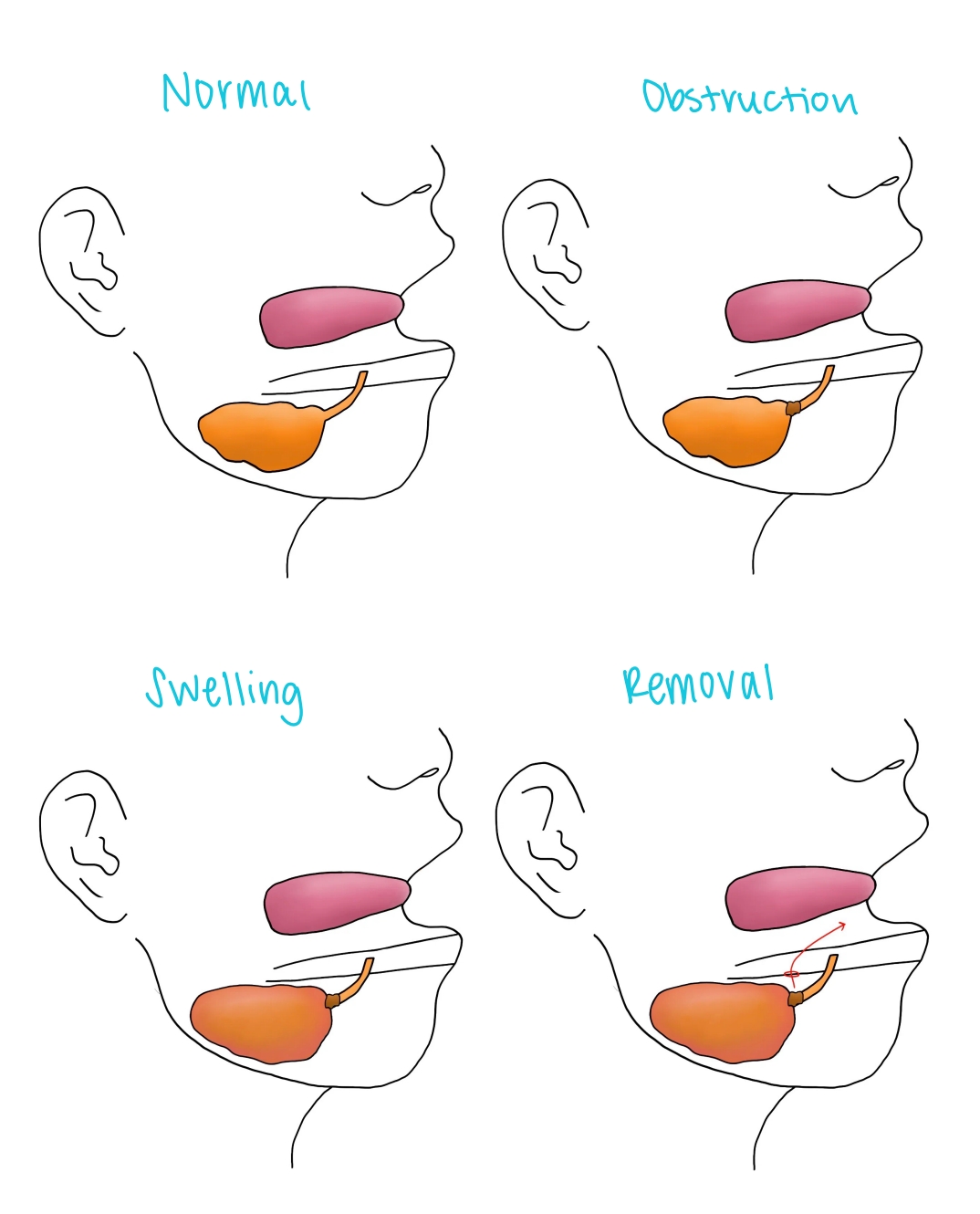
Fellow's annotated case drawing of salivary gland obstruction with the submandibular gland and a calcified stone
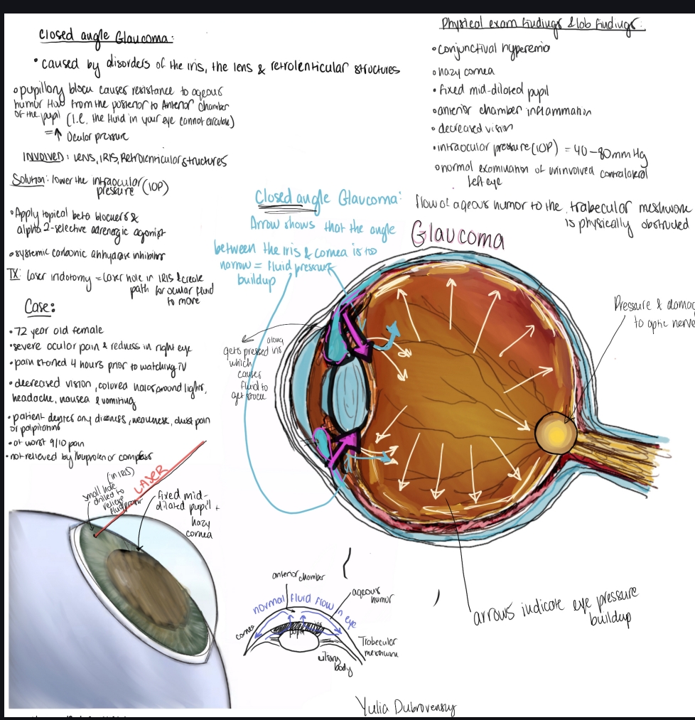
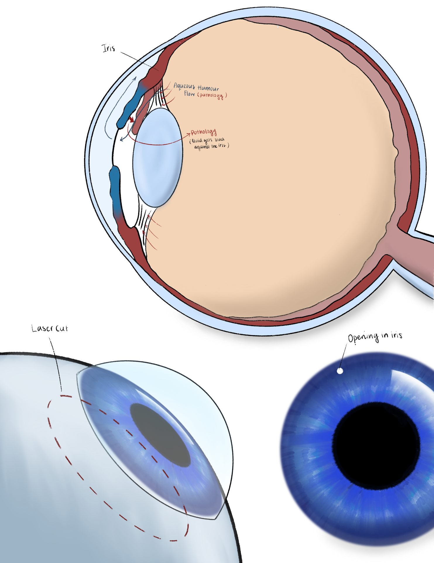
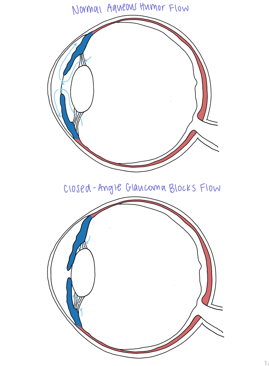
Fellow's annotated case drawing of closed angle glaucoma showing fluid pressure buildup inside the eye
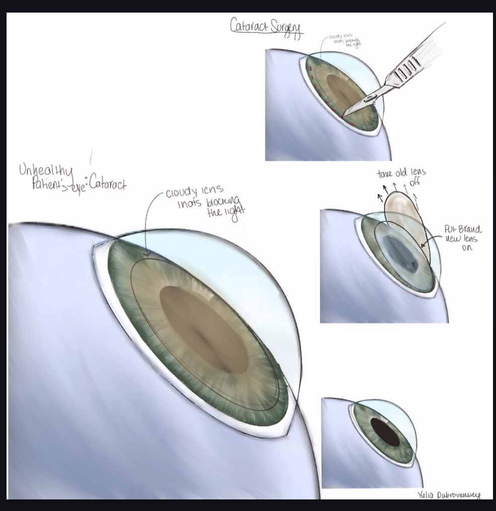
Fellow's multi-panel drawing of cataract surgery on an unhealthy eye with a cloudy lens
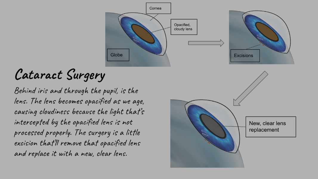
Fellow's labelled explainer of cataract surgery with cornea, opacified lens, excisions and new clear lens
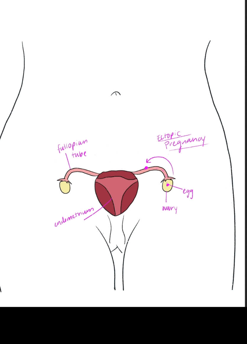
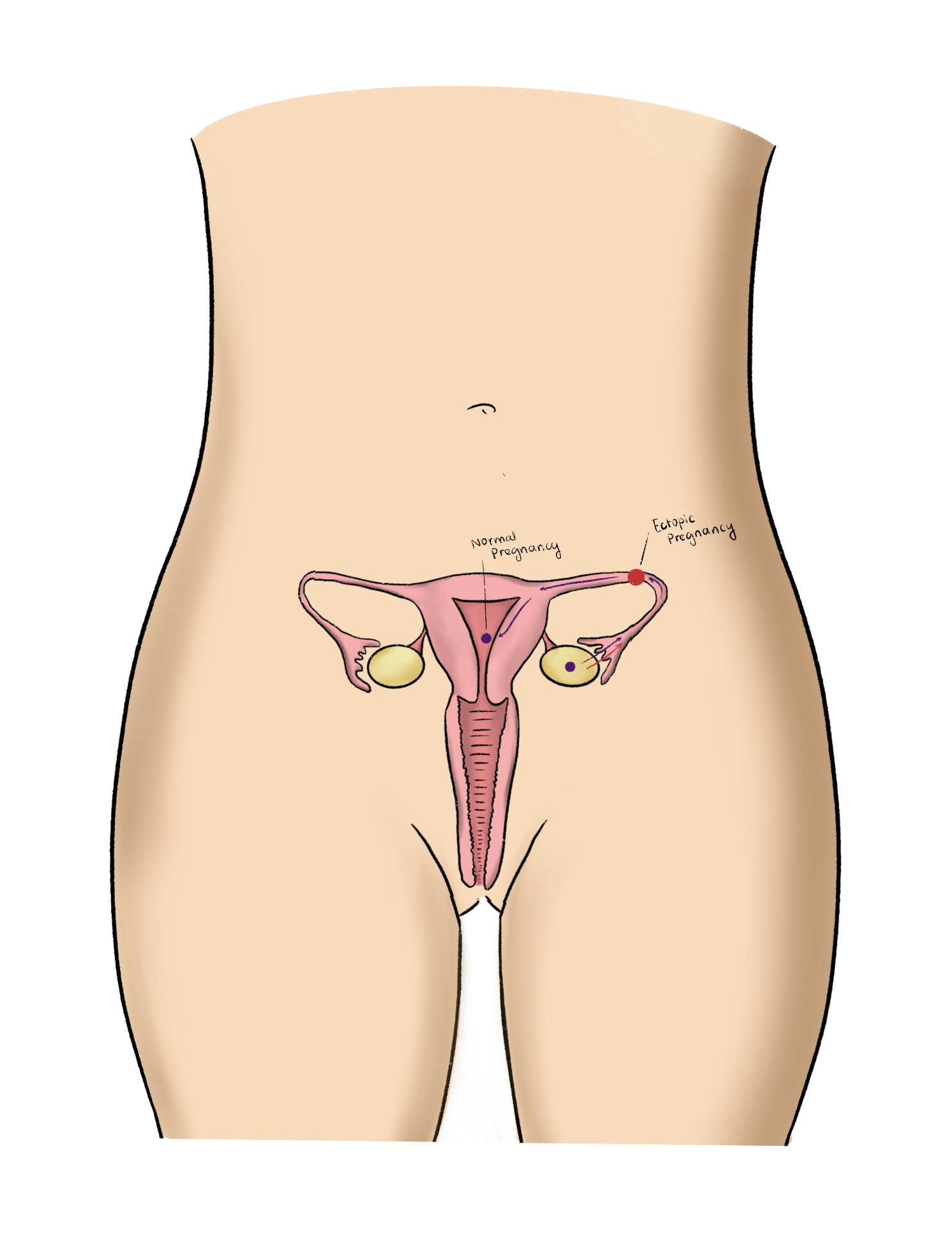
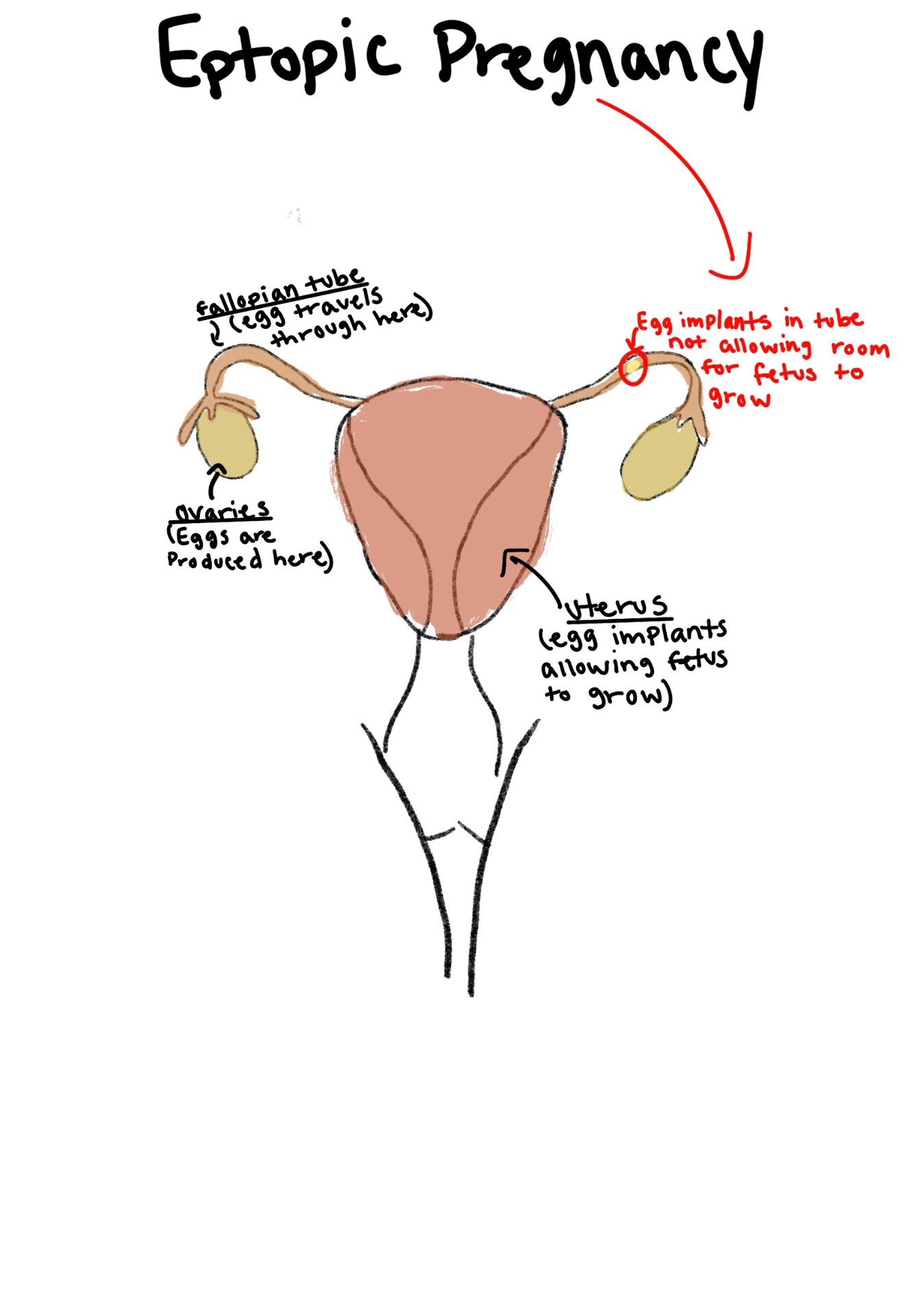
Fellow's drawing of an ectopic pregnancy with labelled fallopian tube, ovary, egg and endometrium
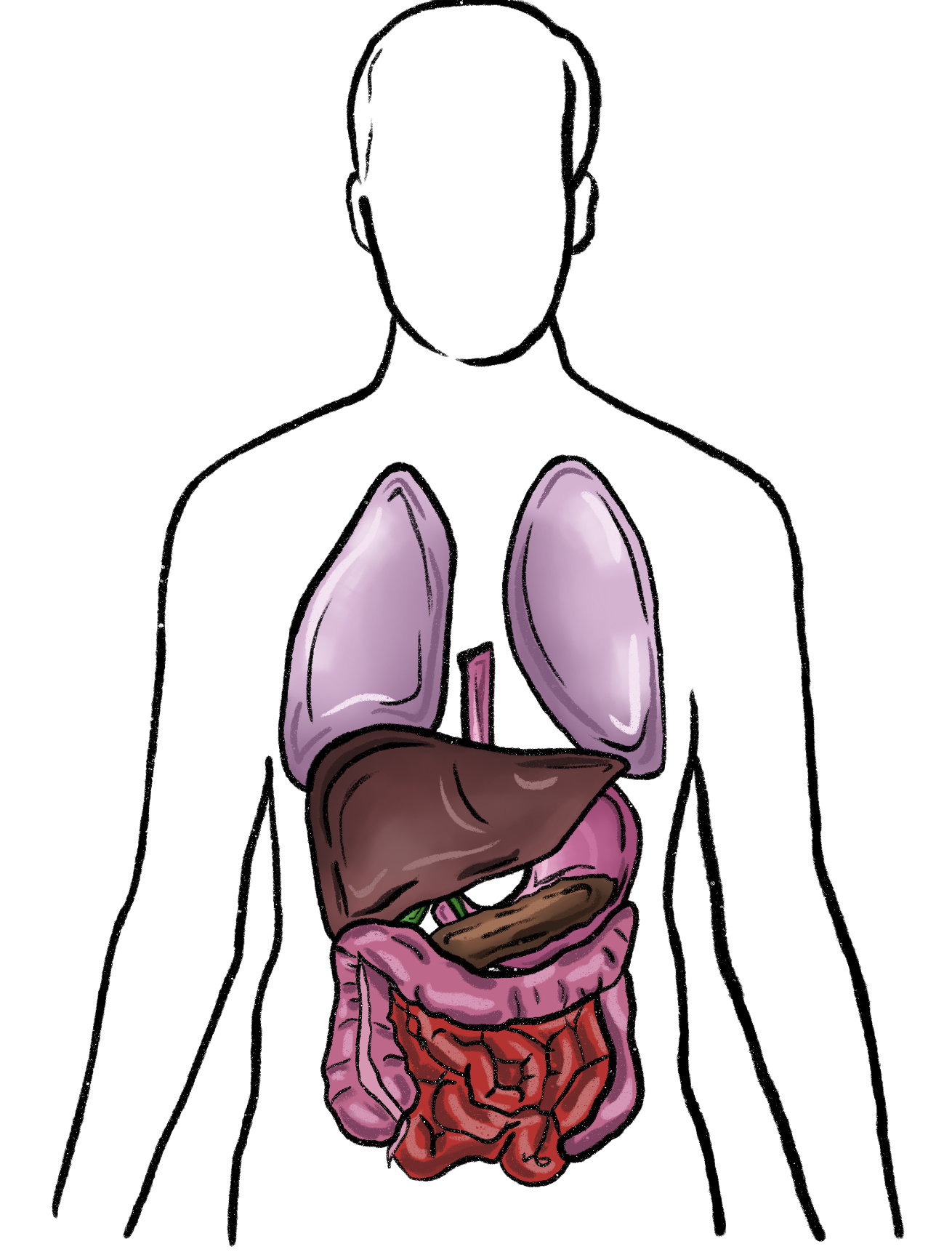
Fellow's drawing of the torso with lungs, liver, stomach, pancreas and intestines
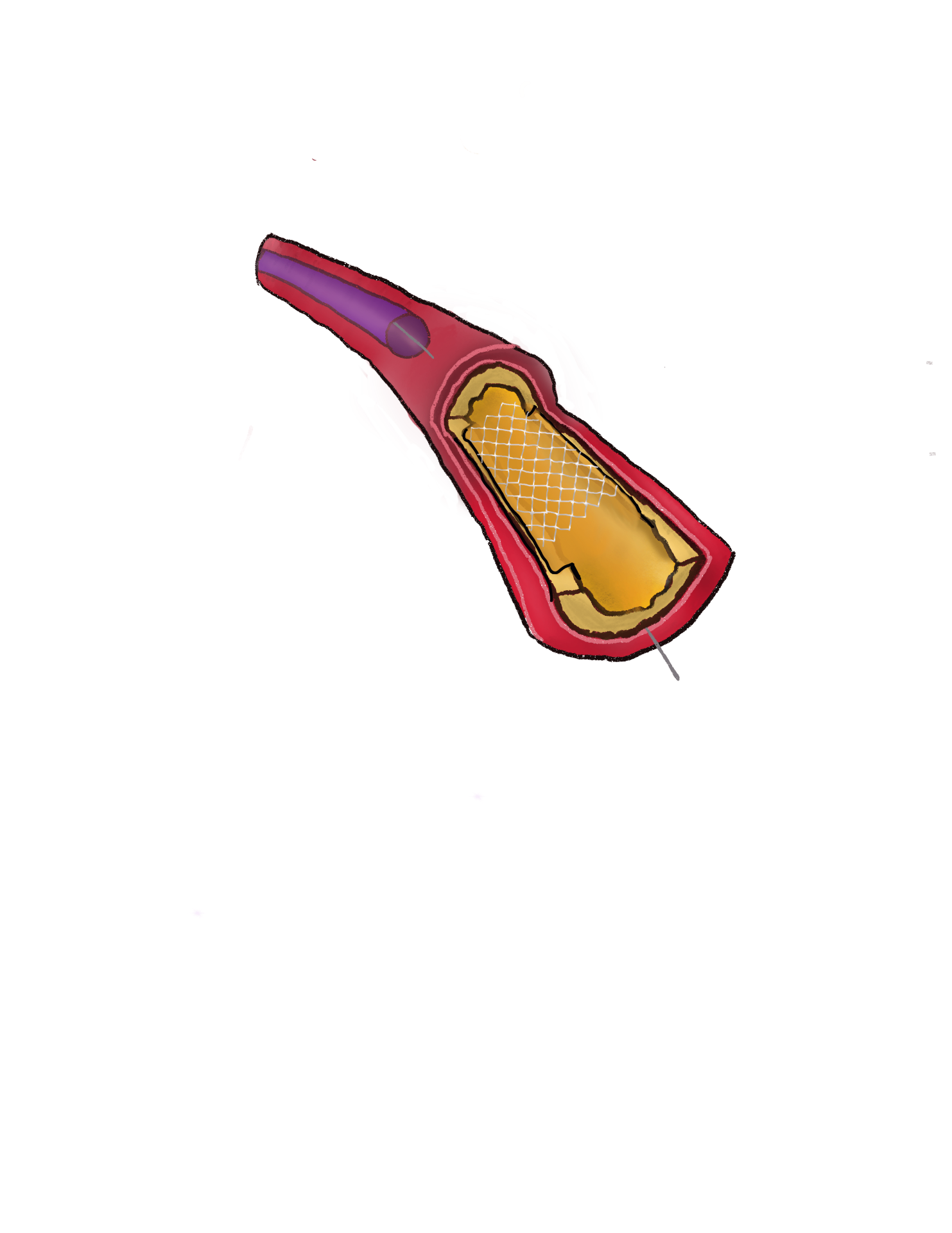
Fellow's drawing of a coronary artery with plaque and an implanted stent
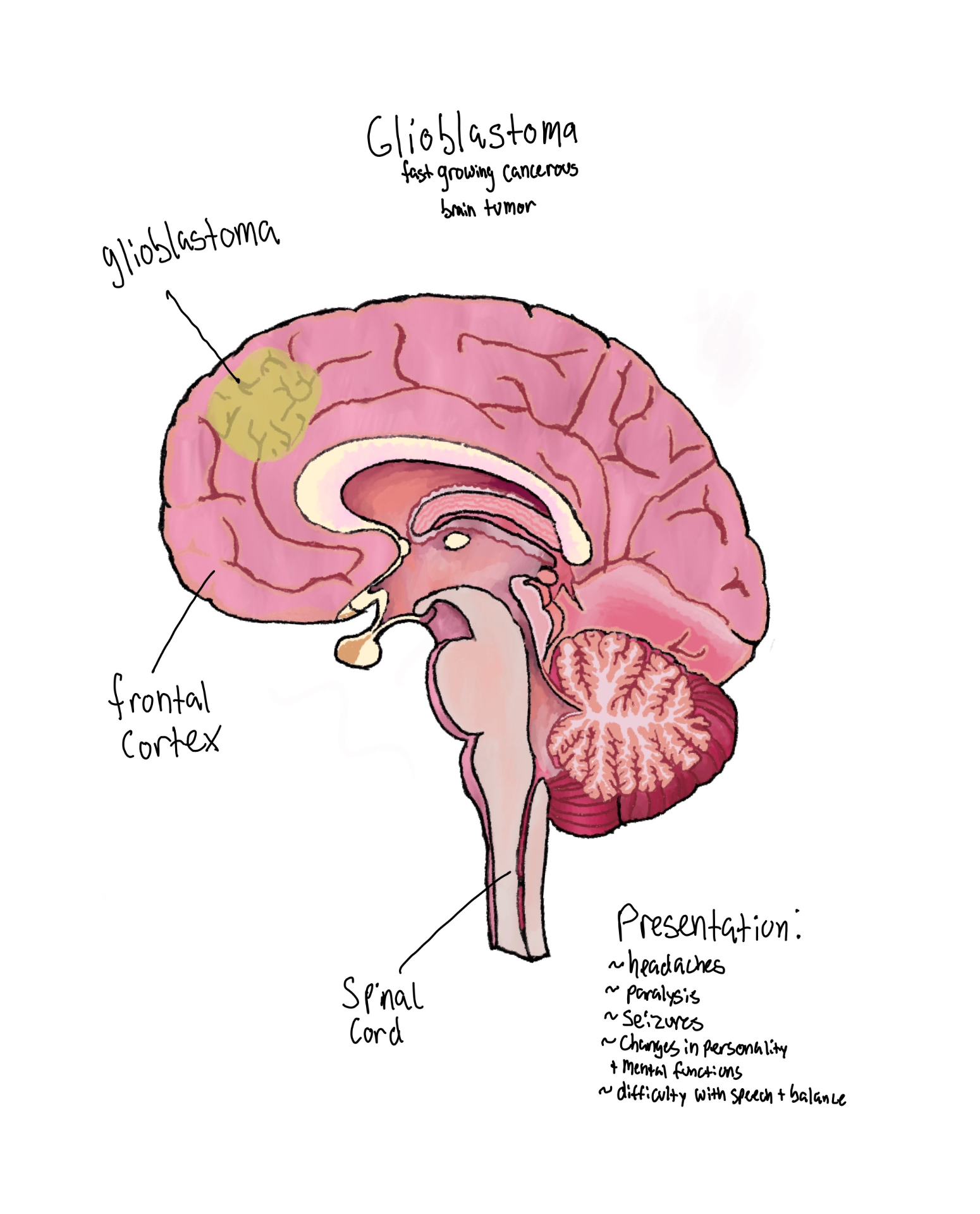
Fellow's labelled drawing of a glioblastoma in the frontal cortex with presentation notes
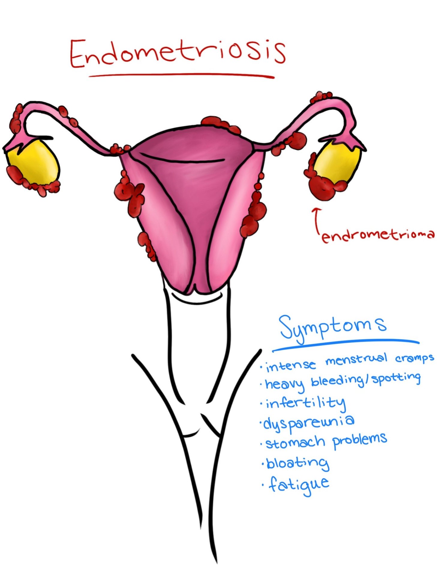
Fellow's labelled drawing of endometriosis with endometrioma and symptom list
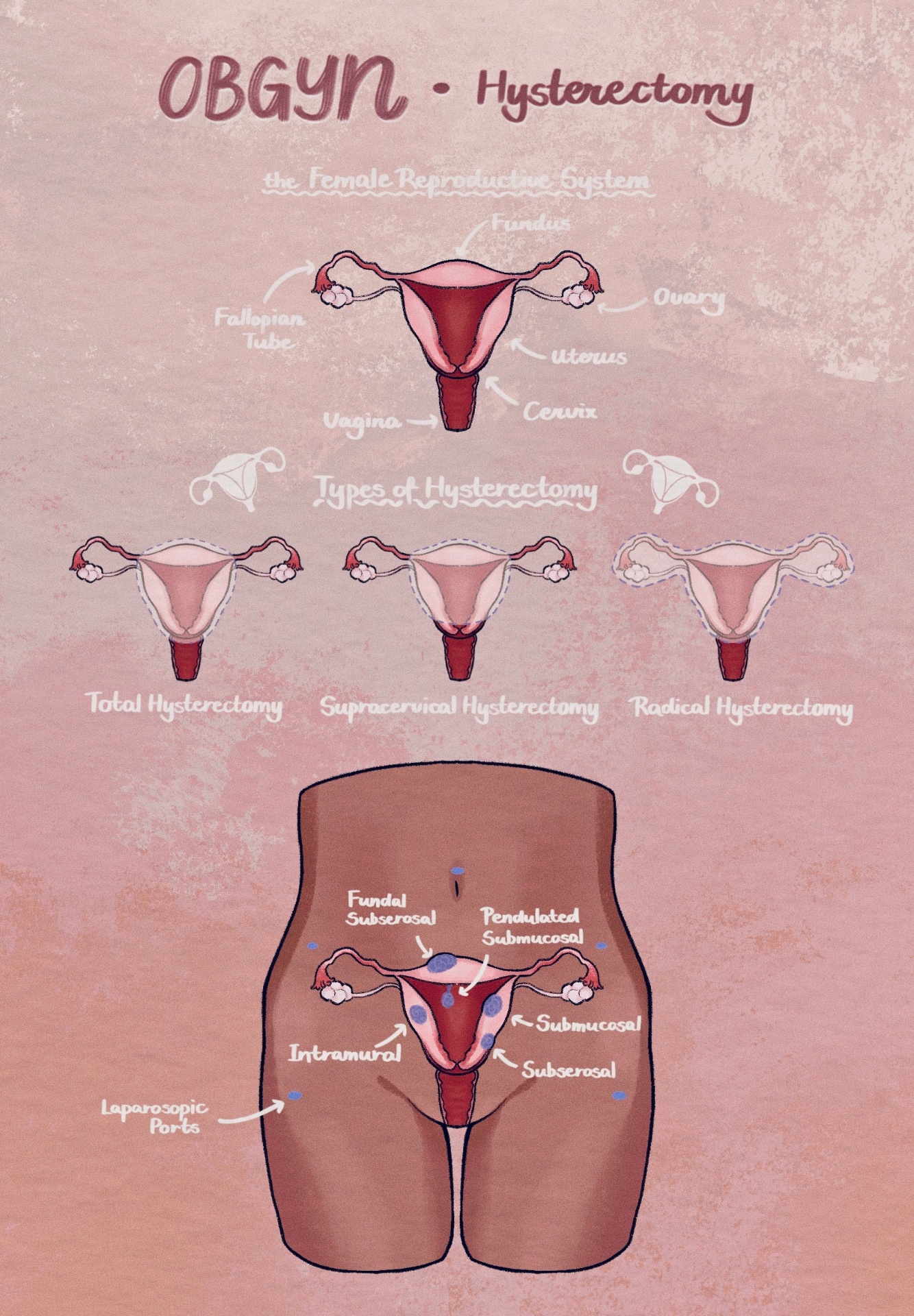
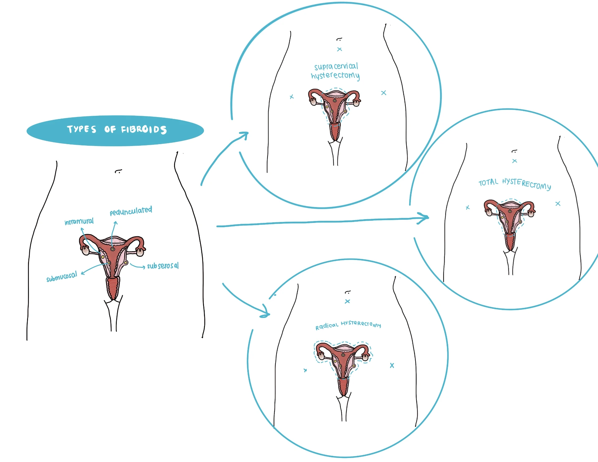
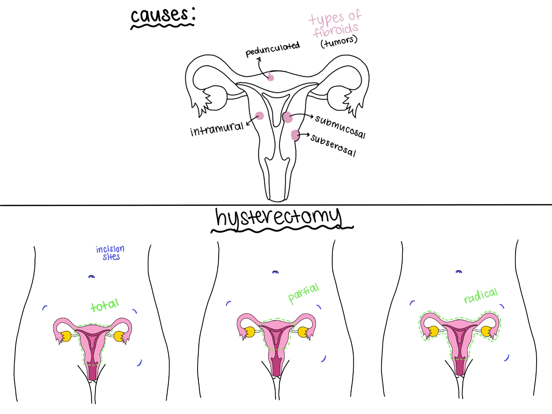
Fellow's illustrated guide to hysterectomy types and the female reproductive system
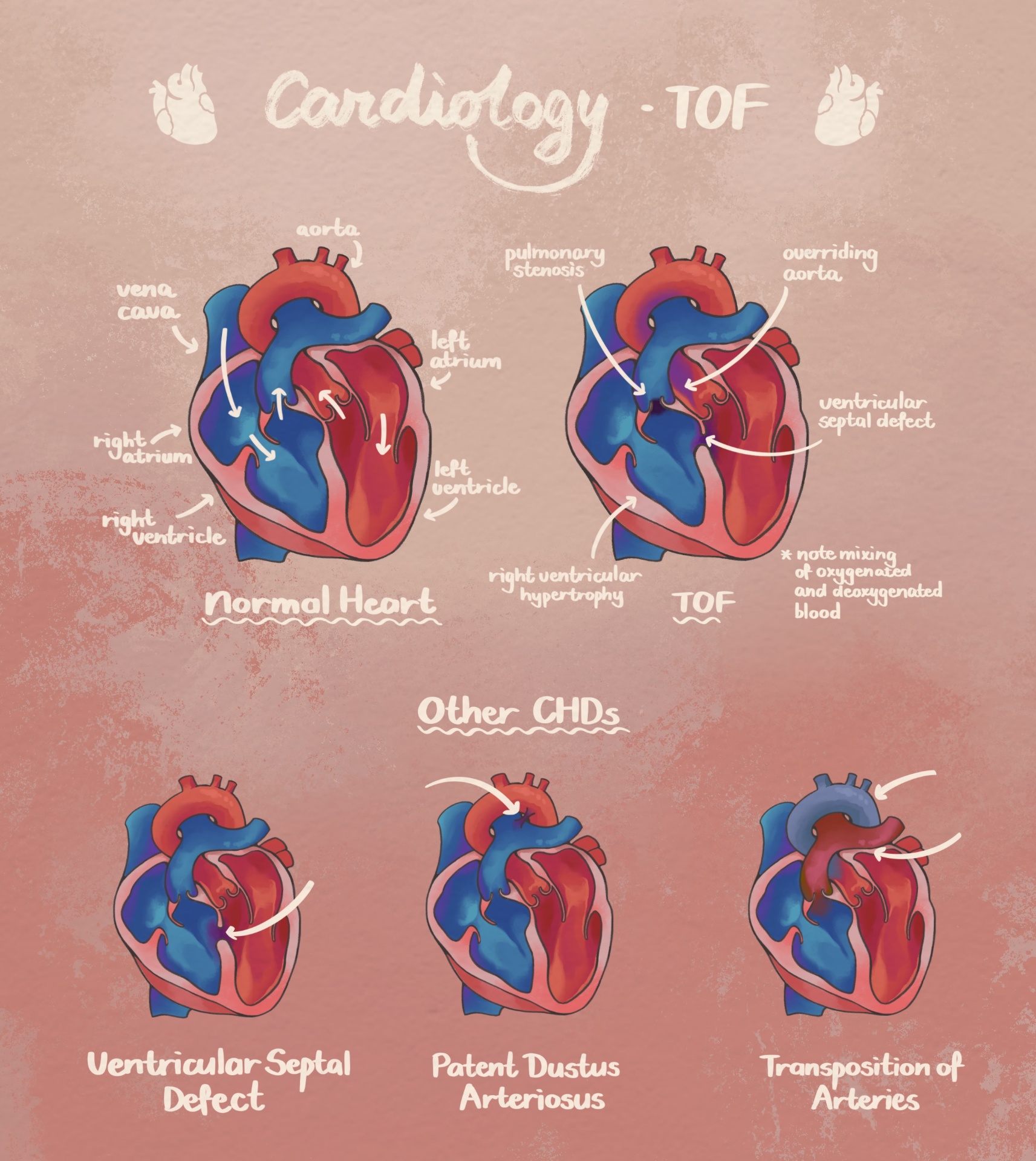
Fellow's illustrated guide to tetralogy of Fallot and other congenital heart defects
Fellow's drawing comparing a normal pregnancy with an ectopic pregnancy in the fallopian tube
Fellow's drawing of aqueous humour flow in glaucoma and a laser iridotomy opening in the iris
Fellow's drawing of a laparoscopic cholecystectomy with clips on the cystic duct
Fellow's drawing of a salivary gland with an obstructed duct in profile view
Fellow's annotated drawing of tetralogy of Fallot with stenosis, overriding aorta and septal defect notes
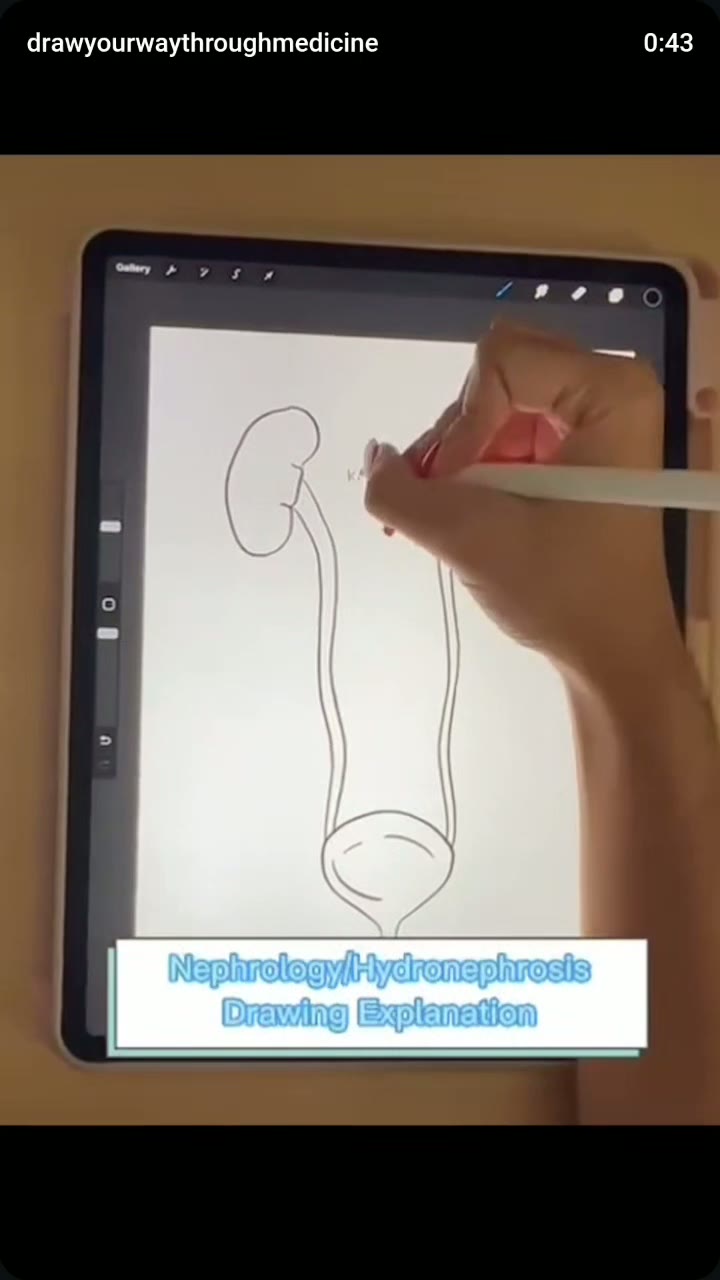
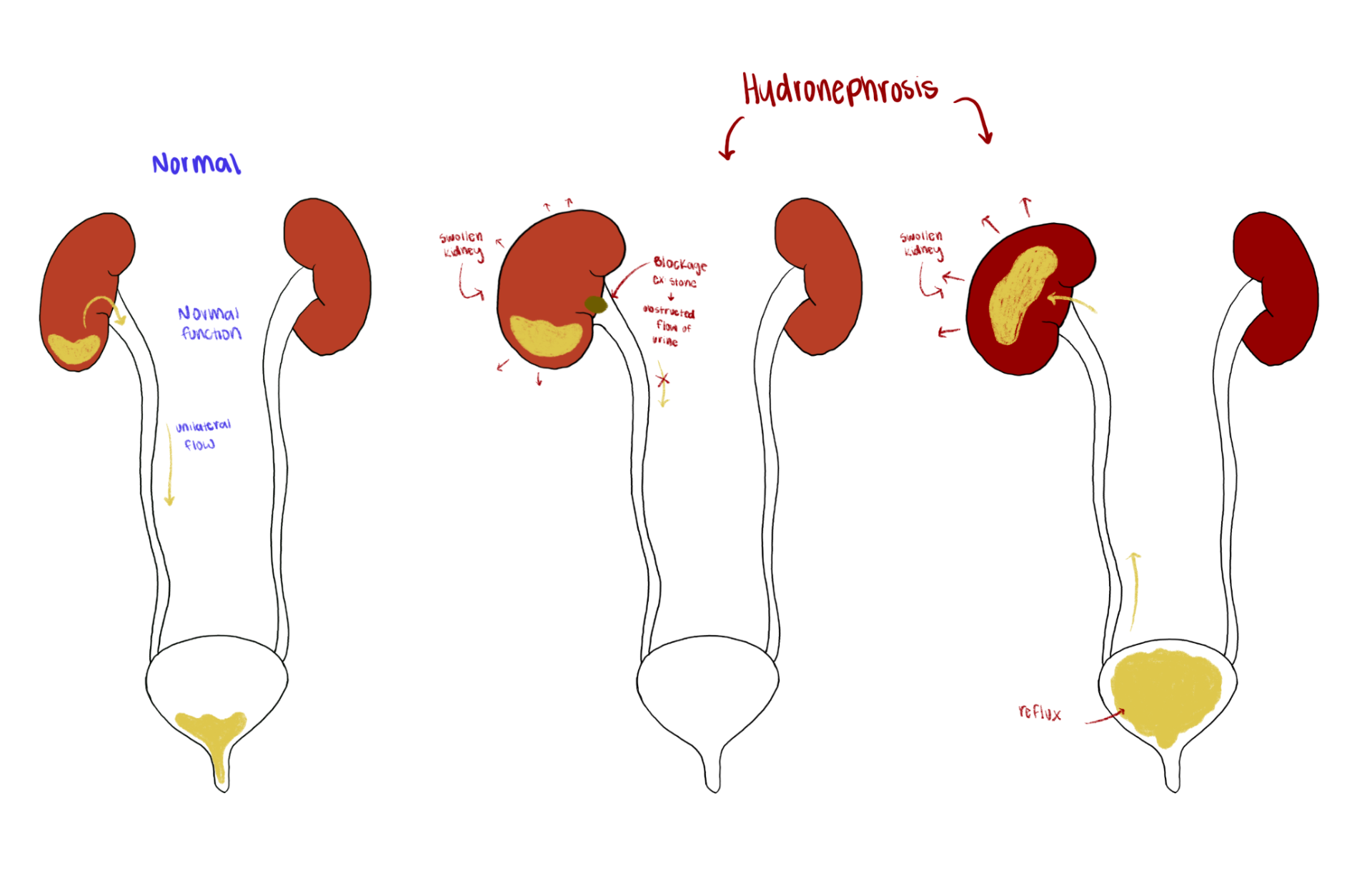
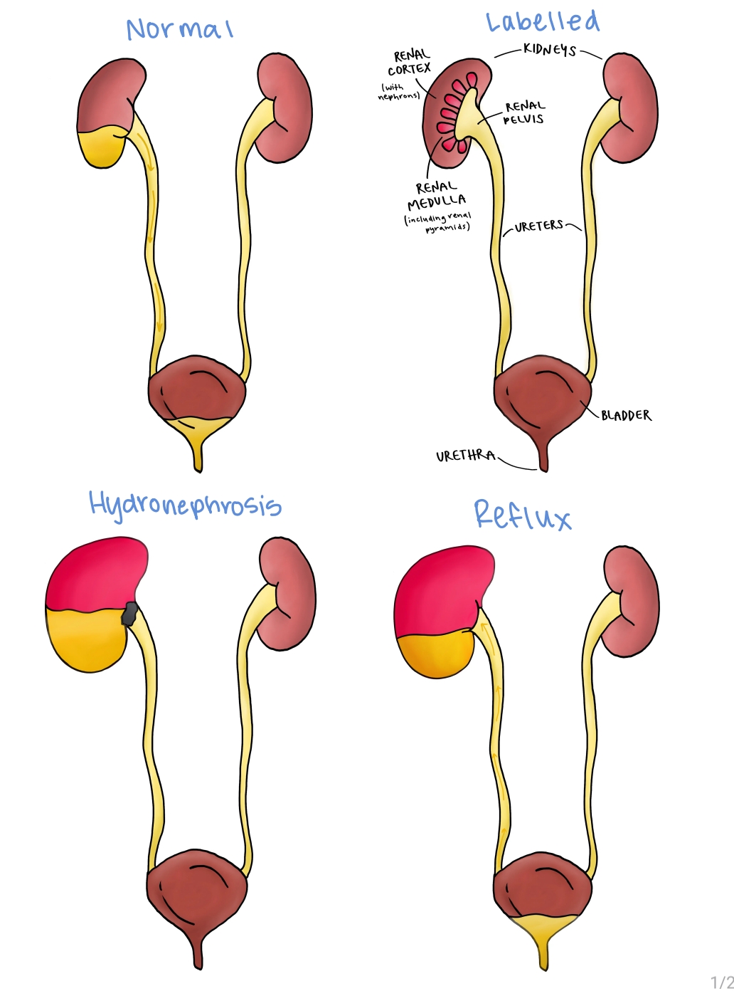
Fellow's drawing comparing normal kidney drainage with hydronephrosis from blockage and reflux
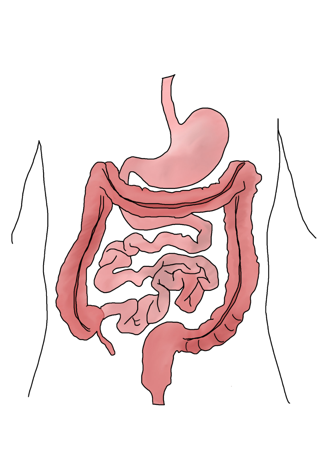
Fellow's drawing of the digestive tract within the torso showing the stomach, small intestine and colon
Fellow's close-up drawing of a mesh stent expanded inside a narrowed artery with plaque
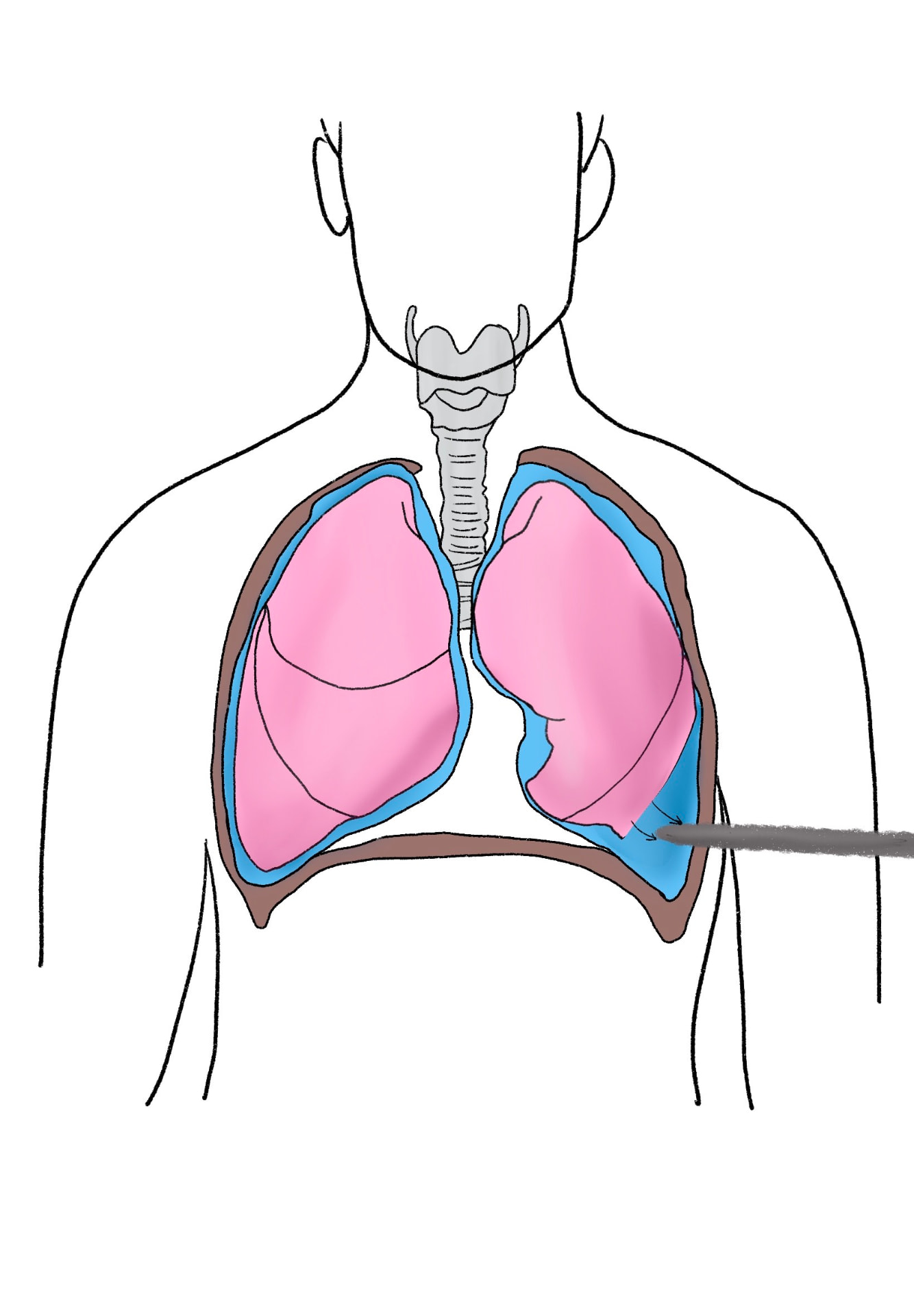
Fellow's drawing of the chest showing lungs, pleural space and a needle draining fluid
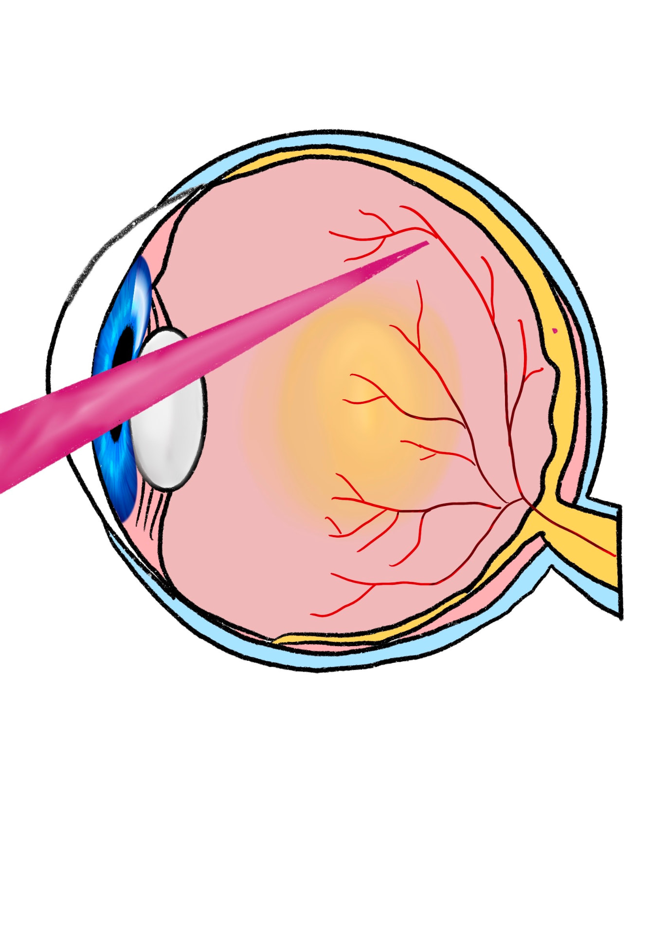
Fellow's cross-section drawing of the eye with a laser beam passing through the lens toward the retina
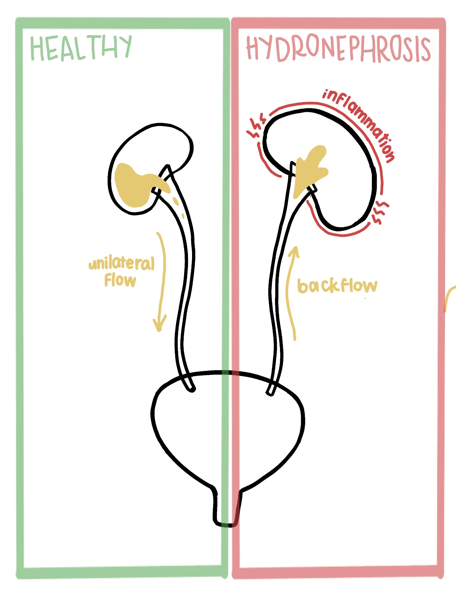
Fellow's side-by-side drawing of a healthy kidney with unilateral flow and hydronephrosis with backflow and inflammation
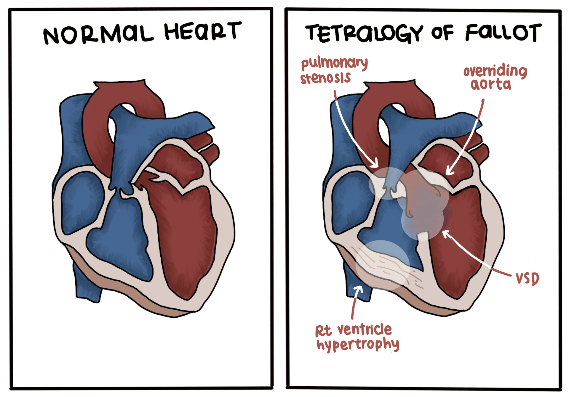
Fellow's side-by-side drawing of a normal heart and tetralogy of Fallot labelling pulmonary stenosis, overriding aorta, VSD and right ventricle hypertrophy
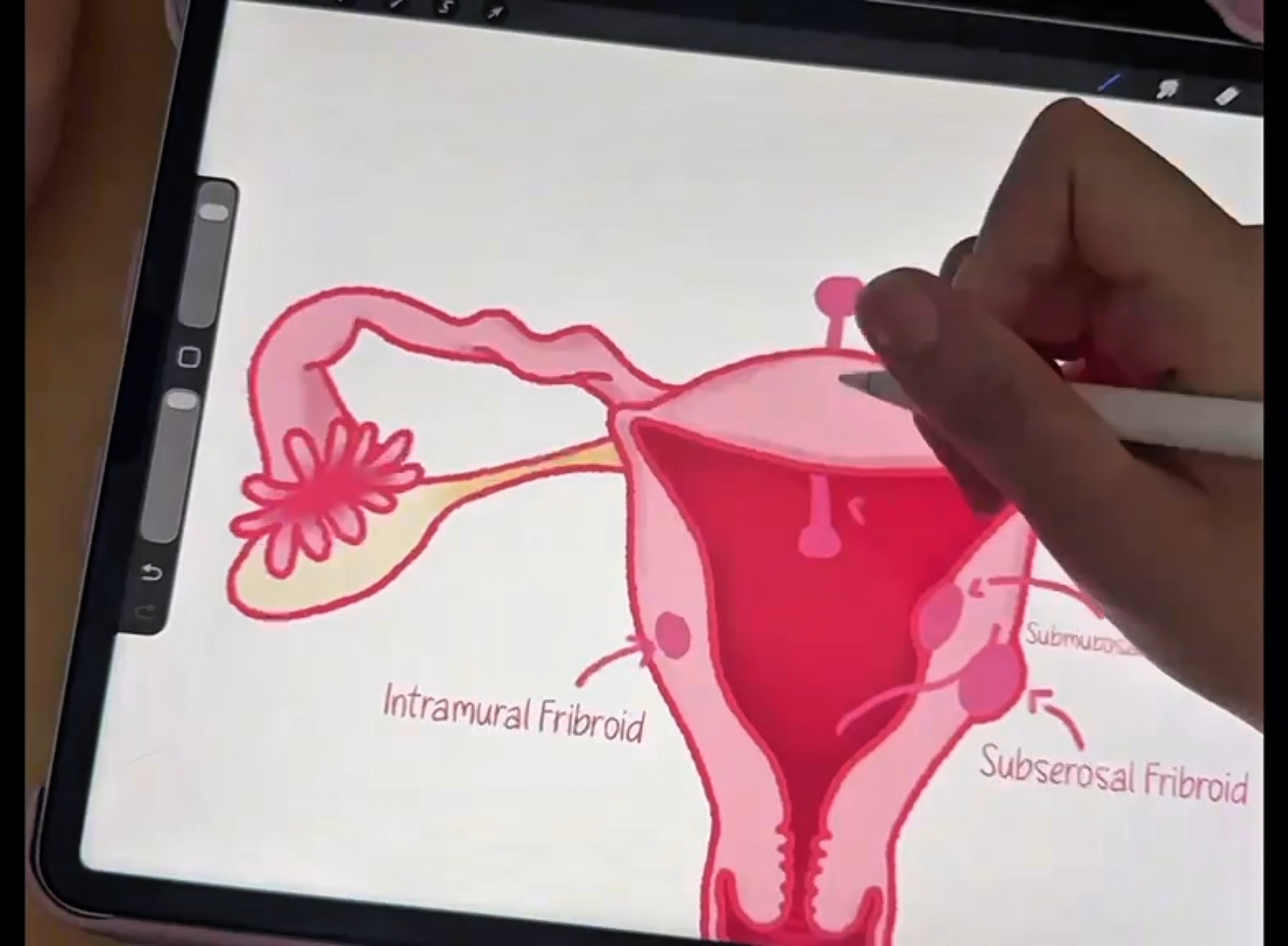
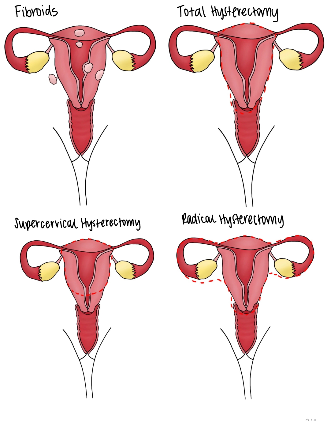
Fellow's diagram of fibroid types and supracervical, total and radical hysterectomy options
Fellow's five-step drawing of a laparoscopic cholecystectomy from clipping to gallbladder removal
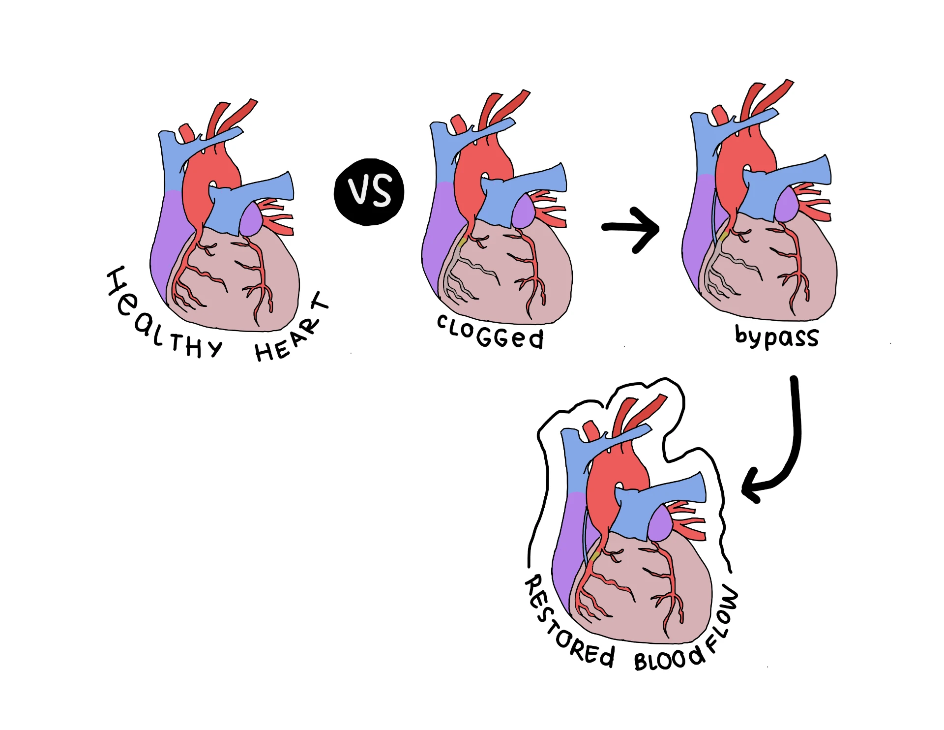
Fellow's drawing comparing a healthy heart, clogged coronary arteries, bypass graft and restored blood flow
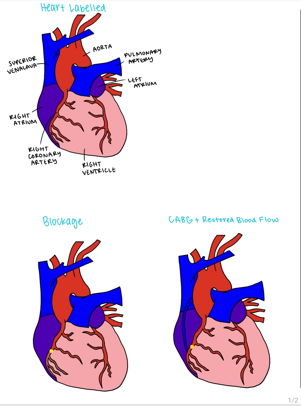
Fellow's labelled heart drawing with aorta, atria and coronary arteries, plus panels showing a blockage and restored blood flow after bypass surgery
Fellow's drawing comparing normal aqueous humour flow in the eye with closed-angle glaucoma blocking flow
Fellow's four-panel profile drawing of a salivary gland showing normal, obstruction, swelling and stone removal
Fellow's four-panel drawing of uterine fibroids with total, supracervical and radical hysterectomy resection lines
Fellow's four-panel drawing of the urinary tract showing normal anatomy, labelled kidney structures, hydronephrosis and reflux
Fellow's multi-panel drawing of the eye showing corneal incision, lens clouding stages and a cross-section of the eye with retinal vessels
Fellow's three-panel drawing of a cholecystectomy showing the liver, gallbladder, clips on the cystic duct and removal
Fellow's drawing of fibroid types in the uterus and total, partial and radical hysterectomy incision sites
Fellow's close-up drawing of cataract surgery labelling the cloudy lens and the corneal incision used to remove it
Fellow's labelled drawing of an ectopic pregnancy with fallopian tube, ovaries and uterus annotations